
I have always taken an interest in the nastier side of life, from keeping pet worms to animal dissection, so it was no surprise that for my degree in Zoology I chose an honours project on poo for my dissertation!
Supervised by Elsa Panciroli (PhD candidate at National Museums Scotland and the University of Edinburgh) and Stig Walsh (Senior Curator of Vertebrate Palaeobiology), I micro-CT scanned carnivore faeces to study the bone fragments of prey encased within them. This novel approach meant that the arrangement and condition of the bone fragments could be studied, which is useful for characterising the faeces of different animals.
My research is part of ongoing work being carried out by National Museums Scotland staff and their research collaborators studying the Kilmalaug Formation, a rock formation that dates from the Middle Jurassic. This is of particular interest as it is one of the few places in the world that contains the fossils of small vertebrates, such as mammals, lizards and salamanders, from this period – a time when we begin to see the first recognisably modern ecosystems.
Researchers want to find out if some of the fossils come from faeces, and if they do, what made them? The animal that made a fossil faeces (or coprolite) is hard to identify because the digestive system varies between different animals. My project aimed to look for ways to identify the producer of a scat by looking at the position of the bones and other fragments of food inside it.
I compared samples of pellets from barn owls and faeces from oriental short-clawed otters and binturong (samples were collected from Edinburgh Zoo). These were packaged up and micro-CT scanned at the University of Edinburgh. I then processed these scans to generate 3D digital models of each sample, showing the bones encased within. Afterwards, I also had the rather messy job of breaking up the samples by hand to check the identification of each individual bone.
To compare the samples, I measured bone volume to matrix ratio, bone erosion, and degree of fragmentation for each bone. I recorded the orientation of bones within the sample by measuring the angle of deviation of the long axis of each bone within each sample from the long axis of the scat. CT scanning let me study all of this before pulling the samples apart, so I could see the bones in situ. I analysed all of this data to see if I could find differences between the three animals.
I expected there would be a difference between the owl pellets and the other two animals, but surprisingly the binturong and otter samples also differed from one another. This is interesting as these animals are very closely related and similar anatomically. The binturong samples contained a lower proportion of bones within each sample, and these varied widely in orientation. In contrast, the otter samples contained the highest proportion of bones and most of them were orientated in the same direction. All animals differed from each other with respect to bone fragmentation (bones within the otter samples were the most fragmented and owl pellet bones were least fragmented), and the bones in owl pellets were the least eroded, while the otter sample bones were most eroded.
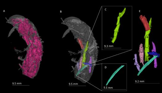
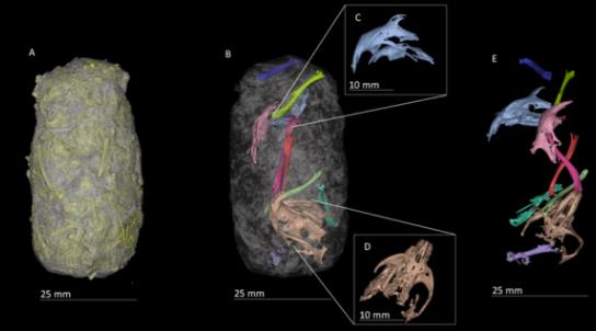
of a birds foot (C), a long fragmented bone (D) and the segmented bones alone (E).
My project was a case study that further research will be built upon. I showed the potential of using CT scanning in the analysis and characterisation of the content of poo – something that had not been done before. My work suggested that animals differ in respect to the arrangement and modifications of bones within their poo. This is important as it could be used as a diagnostic tool to identify fossilised poo samples, which is currently very difficult.
I thoroughly enjoyed my research project and went on to achieve a first in my dissertation. This project solidified my passion for research and practical work, which I plan to carry forward into a research position and further study.
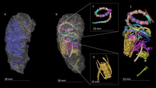
All 3D images created and captured in Mimics Research 19.0 by Materialise.